
Mastering “The Mystic Mastoid” Part 3
The Key to Surgical Excellence
Introduction of the methodology
The blogs written under the aegis of OssaPlus Simulations, the makers of 3D models of various bones and organs, will cover the various subjects, e.g. why simulation workshops, what is the importance of artificial models, how to approach and dissect a specific artificial model, etc. Readers are at free will to interact with the author through the contact page about the articles written in the blogs.
Every organ in our body is well protected in its own way.
The mastoid bone, a part of our temporal bone, fuses with the petrous part of the bone in our early foetal life. This petromastoid portion, a very tough bone, holds the organ of hearing and balance. More so, this organ is very closely related to other very important structures like internal carotid artery, facial nerve, sigmoid sinus, dura, etc. The arrangement is so compact and intricate that reading books or seeing the diagrams can never give the three-dimensional perception of their anatomical layout. Leave apart doing live surgery on this bone.
Mystic means full of mysteries, nothing to do with mysticism. Those mysteries make every mastoid bone unique. Even the bones of either side are not exact replica of each other. To gain the first-hand knowledge of its mysteries, there is no other way than to dissect the multiple versions of this bone.
Undeniably, surgery is an art, a very fine art. More so in the case of mastoid surgeries, and even more if surgeon wants to access structures beyond mastoid. Going via the route of labyrinth for the neurotological access of deeper lesions needs super skills and dexterity of handling the instruments. Just like a painter, slightest touch of whose brush at wrong spot can damage the whole work, slightest touch of a running drill at wrong spot can play a great havoc with the surgery.
There is no doubt that a teacher shapes a student. A good experienced mentor, who can teach you the correct ways of handling the instruments, the right paths to the deeper structures, the care to be taken while handling the delicate soft tissues, the ideal methods of tackling the complications, and all the underlying philosophical thoughts behind every surgical method, is a very essential asset. In addition, as any singer will agree, regular daily practice of singing is required to correctly hone the singing apparatus. Even a skilled otologist needs regular practice of temporal bone dissection. Each time he will find new surprises in this bone. While dissecting, minute attention to the variations of colour, the change of sound as the drill burr touches the underlying bone, the haptic feel of the hardness or softness of the drilled tissue, the interrelations of the compactly arranged vessels and nerves, and the acute vision like an eagle, all these are to be learnt a hard way. By repeated dissections. Then only can you be a fairly skilled mastoid surgeon.
Even a skilled otologist needs regular practice of temporal bone dissection. Each time he will find new surprises in this bone.
Though dissection of cadaveric bones is the most ideal simulation exercise for training hands, it is just impossible to get such versions of cadaveric bones. Hence the need of artificial temporal bones.
The search for alternative to cadaveric bones started with the acute paucity of the bones. Ethical, legal, religious, contagious, and many such issues arise with the use of cadaveric bones. Virtual temporal bone dissection methods are already in use, some giving the feel of bone different than soft tissues, and even blood oozing and colour variations of tissues. But, all said, they can never give the exact haptic feel and hence cannot fully replace the actual dissection of the bone. They are on screen, not on actual three dimensional model.
Various attempts to make the artificial bones are reported in literature. Some are just sufficient to teach the external anatomy, some are meant for hands on dissections. Obviously, there are a multitude of challenges in making the artificial bones. Right from getting the correct materials for building such bones to replicating the correct topological layout of all those minute anatomical details is a great task. Additionally, giving the exact haptic feel of the structures during dissection practice must be achieved. Every channel or canal in the bone should be patent. Otherwise, dissecting bones with incorrect haptic sensations will build up a false confidence in the dissector.
The writer of this blog uses the 3D printing technology to make the temporal bone and then its meticulous manual crafting gives the final bone, complete with colour codes and soft tissues and nerves and vessels, etc. The composite material used in this bone is a unique blend of different nontoxic chemicals and resins. The mix has been optimised to attain the near hardness and consistency of a cadaveric bone.
This artificial temporal bone, made out of the CT scans of the temporal bone of a volunteer, is unique in that it has all the external as well as internal anatomical features, exactly placed. All the semicircular canals and the cochlea are patent. All the nerve canals are holding the nerves. Seventh cranial nerve, chorda tympani, and greater superficial petrosal nerve are present. Internal carotid artery as well as sigmoid sinus are in their places. Pink dura is very discernible. Most importantly, the complete ossicular chain is in its correct position. As the printing progresses at the 50-micron level, every minute detail has been replicated here.
Straight from the bench, unretouched: the printed bone before it reaches a delegate’s drill.
On this model of temporal bone, anything from simple mastoidectomy to translabyrinthine approach as well as cochlear implant surgery can be learnt. Dissection of one such bone can give the experience of more than twelve different procedures, if gone stepwise. The technology has enabled us to make different types of mastoid bones, say adult, paediatric, differently pneumatized, diseased, congenitally anomalous, etc. Some bone models can be exclusively for learning the stapedotomy surgery, others only for ossiculoplasty or tympanoplasty. There can be a glomus tumour or cholesteatoma in some, and others may be only for advanced cochlear implant dissection.
Any surgeon can send the DICOM files of CT scans of their patients and we can make the bone from it.
To do a mock surgery on your patient's mastoid bone, to have the full idea about what lies inside, to practice tackling the proposed complications, and all such, make a huge difference in building the surgical confidence of a surgeon. Fortunately, all this is possible with this technology of 3D printing and artifical bones.
Though many models of artificial temporal bones are available, only a few have all the external as well as internal details of this bone. The unique feature of the bones used by the writer of this blog for teaching the hands-on dissection is that it gives the anatomy landmarks and has the experimentally measured haptic feel of the bone. These bones, after making, are tested at four levels, viz., reading the external anatomical features of the bone, comparing the drill response to the cadaveric bones, comparing the CT scans of these bones with those of the DICOM files from which they are made, and taking the validation of them from reputed otologic surgeons. Each bone undergoes the strict quality check before being handed over for the hands-on practice.
It is even possible to have various in-built sensors in the bone for the facial nerve or dura or sigmoid sinus. Though not incorporated in each bone, the sensors alarm the dissector about the accidental breach or injury of the important structures. The details of these will be discussed in further blogs.
As is well known, the field of 3D printing is very vast and fast developing. New materials and new techniques arise fast. The writer had been using different printing methods previously, which had their flaws. The heat generated by the drill used to melt the material and deform the underlying structures. The recent new version of this bone, made from the composite that is under the patent process, gives the exact bone dust while drilling. This can be irrigated and sucked out as in the cadaveric bones.
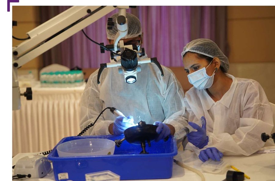
All the normal mastoid surgery instruments can be used during the hands-on dissection on this model. The plus point is no fear of disease transfer and the absence of the need of any preservative. There are no legal or religious issues associated with this.
All said, each artificial model comes with a certain shelf life. Beyond that life, the model, though dissectible, will lose marginally on the exact haptic feel and will develop some micro fractures in its outer shell. Though no special preservation is needed, it is advisable to store these bones so that they are not exposed to extreme temperature variations. And after dissecting, advisable to discard them in a proper way, like burying them or burning them. Also, during dissection, taking the basic precautions as wearing a gown, gloves, cap, mask and eye-protection is advisable. Keep in mind that constant irrigation is a must. Dry run of the drill on this bone for a long duration can give a burning smell. And the fumes generated prove noxious. Apart from the bone dust, the soft tissues in this bone use various types of silicone or latex and such. Hence strictly avoiding any accidental ingestion of these materials is very essential. Similarly, drilling done without proper precaution can generate minute fractured pieces of the bone and their accidental landing on the cornea needs to be avoided. That is why suggested that all the precautions, as taken during live surgery, are a prerequisite for the hands-on dissection of these models.
What lies ahead, in the next two parts of this blog, Mastering the Mystic Mastoid, is the discussion of anatomy details present in this bone and the in-depth description of the various procedures to be done during its dissection. If followed in a certain protocol, the dissector will have a certain amount of basic confidence to do the live surgeries.
In addition to the temporal bone, there will be discussions about other artificial models, as of nose, larynx, salivary glands, vestibule, etc.
Beyond the temporal bone: the rhinology model, with a lifelike external nose over the full sinonasal anatomy.
Train on this bone
The models and programmes described above, at ProGuide.
- The temporal bone models— basic, complete and task-based
- Your own model from a CT scan— send the DICOM files, as described above
- KBI SkillBridge workshops— dates, fees and registration
- Ask a question— our programme experts answer seven days a week
Writer
Dr Prashant M Naik
MS (ENT), DLO
Nagpur, India